Side View of the Skull ClipArt ETC
Browse 1,897 human skull side photos and images available, or start a new search to explore more photos and images. NEXT Browse Getty Images' premium collection of high-quality, authentic Human Skull Side stock photos, royalty-free images, and pictures. Human Skull Side stock photos are available in a variety of sizes and formats to fit your needs.

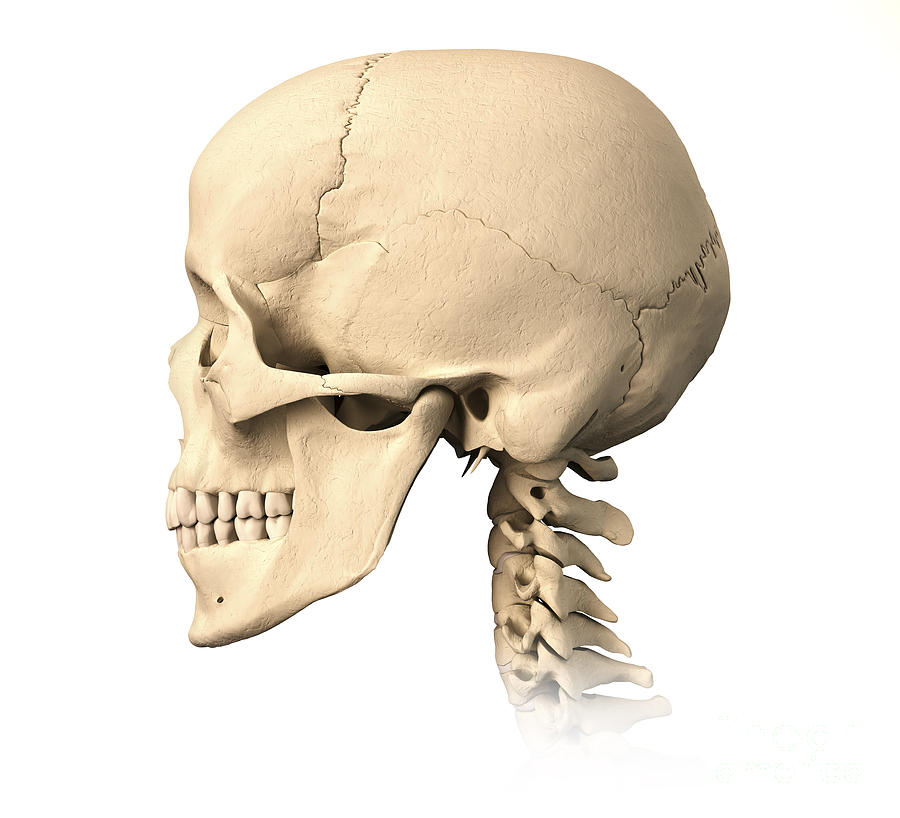
Side View of an Antique Human Skull Isolated on White Stock Image Image of natural, body
Parts Conditions Treatment The occipital bone is a flat, trapezoid-shaped bone that houses the back part of the brain. It is located at the lower back of the cranium and is one of seven bones that form your skull. This article will review the structure and function of the occipital bone of the skull, as well as problems that can affect the bone.
Skull anatomy Anterior and lateral views of the skull Kenhub
The skull, also known as the cranium, is the group of bones that forms the head. While many people think of the skull as a single structure, it's actually made up of 22 bones that include the.
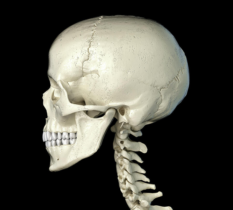
Anatomy Of Human Skull, Side View Photograph by Leonello Calvetti
What is occipital neuralgia? Most feeling in the back and top of the head is transmitted to the brain by the two greater occipital nerves. There is one nerve on each side of the head.

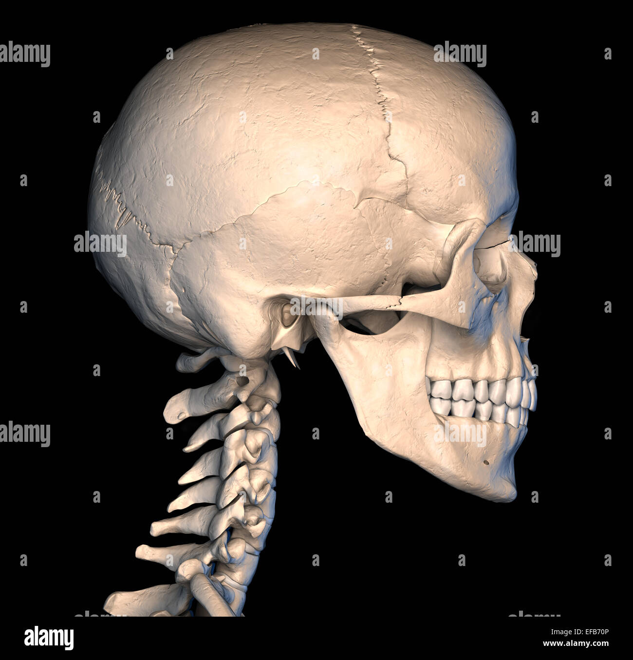
lateral view of skull Simon Hart
External Website Watch this video to view a rotating and exploded skull, with color-coded bones. Which bone (yellow) is centrally located and joins with most of the other bones of the skull? Anterior View of Skull
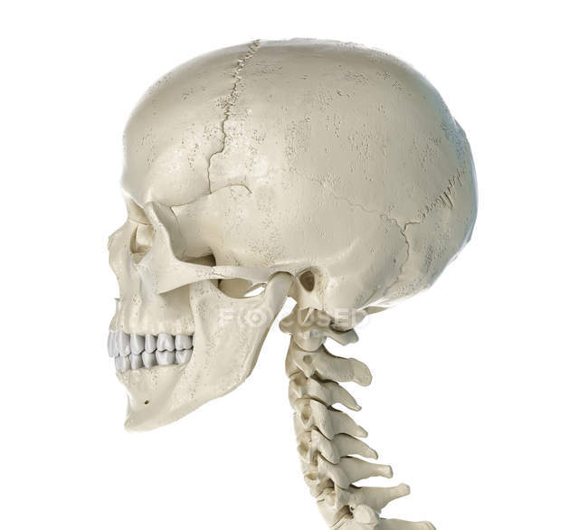
Side Profile Of The Human Skull Photograph by Leonello Calvetti Pixels
Skull, skeletal framework of the head of vertebrates, composed of bones or cartilage, which form a unit that protects the brain and some sense organs. The skull includes the upper jaw and the cranium.. The atlas turns on the next-lower vertebra, the axis, to allow for side-to-side motion. inferior view of the human skull. internal surface of.
Human skull in side view on white background. — medical, cranium Stock Photo 275202828
On the base of the skull, the occipital bone contains the large opening of the foramen magnum, which allows for passage of the spinal cord as it exits the skull. On either side of the foramen magnum is an oval-shaped occipital condyle. These condyles form joints with the first cervical vertebra and thus support the skull on top of the vertebral.
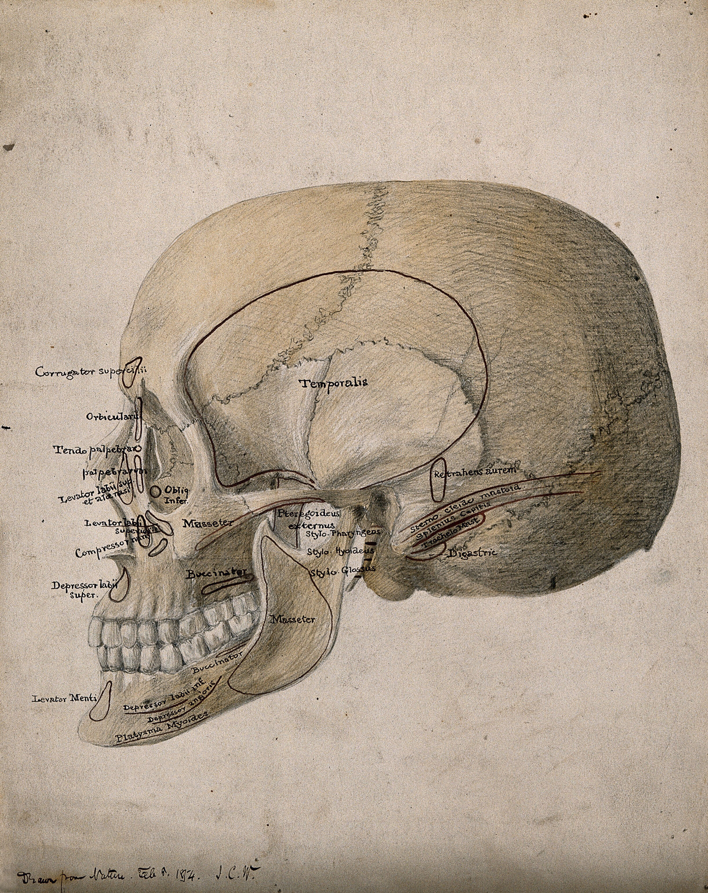
Human skull side view. Watercolour, ink and pencil drawing by J.C. Whishaw, ca. 1854
Humans Skull in situ Anatomy of a flat bone - the periosteum of the neurocranium is known as the pericranium Human skull from the front Side bones of skull The human skull is the bone structure that forms the head in the human skeleton. It supports the structures of the face and forms a cavity for the brain.
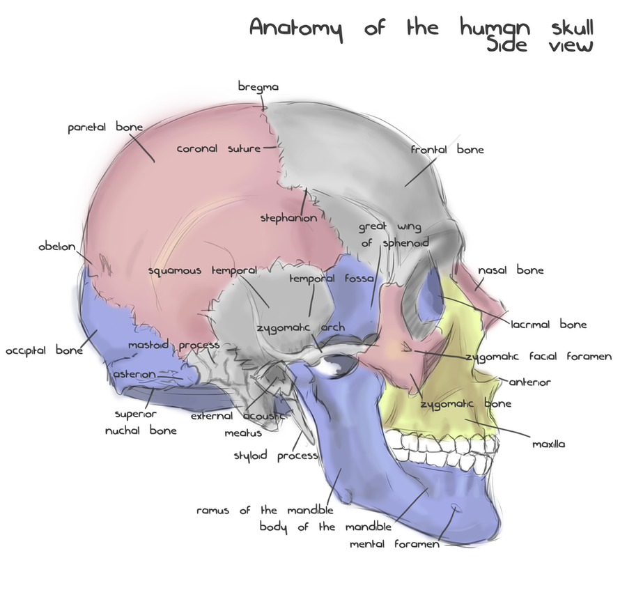
Annotated human skull anatomy side view by shevans on DeviantArt
Side view. On black background. Human Skull, SideView, Skeleton Head, Clipart , Vector Illustration Human skull in different angles. Isolated on black background. Side and front views. Anatomy and medicine concept. Vector isolated one single simplest smiling skull dead head isometric side view colorless black and white contour line easy drawing
Skull Human Side View White Background High Resolution Stock Photography and Images Alamy
1/2 Synonyms: none The human skull consists of 22 bones (or 29, including the inner ear bones and hyoid bone) which are mostly connected together by ossified joints, so called sutures. The skull is divided into the braincase ( neurocr anium) and the facial skeleton ( viscerocranium ).
Introduction Anatomy and Physiology
Symptoms Diagnosis Treatment Recovery and long-term effects Takeaway The underlying cause of a skull fracture is a head trauma significant enough to break at least one bone. People with a.

Skull side view Diagram Quizlet
1/20 Synonyms: none The posterior and lateral views of the skull show us important bones that maintain the integrity of the skull. The posterior surface protects the region of the brain that contains the occipital lobes and cerebellum .
7.2 The Skull Douglas College Human Anatomy and Physiology I (1st ed.)
Anatomy and medicine concept. Human Anatomy full body male skeleton. Five views. Perspective, Front rear and side on black background. 3d illustration. Realistic human skulls front and side views set isolated on white background vector illustration Set of realistic skeletons isolated on gray background. Anterior, lateral and posterior view.

Adult human skull. Side view Xray showing the cranium (Photos Framed Prints...) 6420405
The skull contains all the bones of the head and is a shell for the brain and the origins of the central nervous system. A first glance shows that this is one large mass of detailed and irregular bone. Upon closer inspection however, it seems that it is intricately constructed of many smaller bone fragment pairs, all unique in shapes and sizes, that bilaterally make up this hollow, three.

Skull Side View Horror Free vector graphic on Pixabay Pixabay
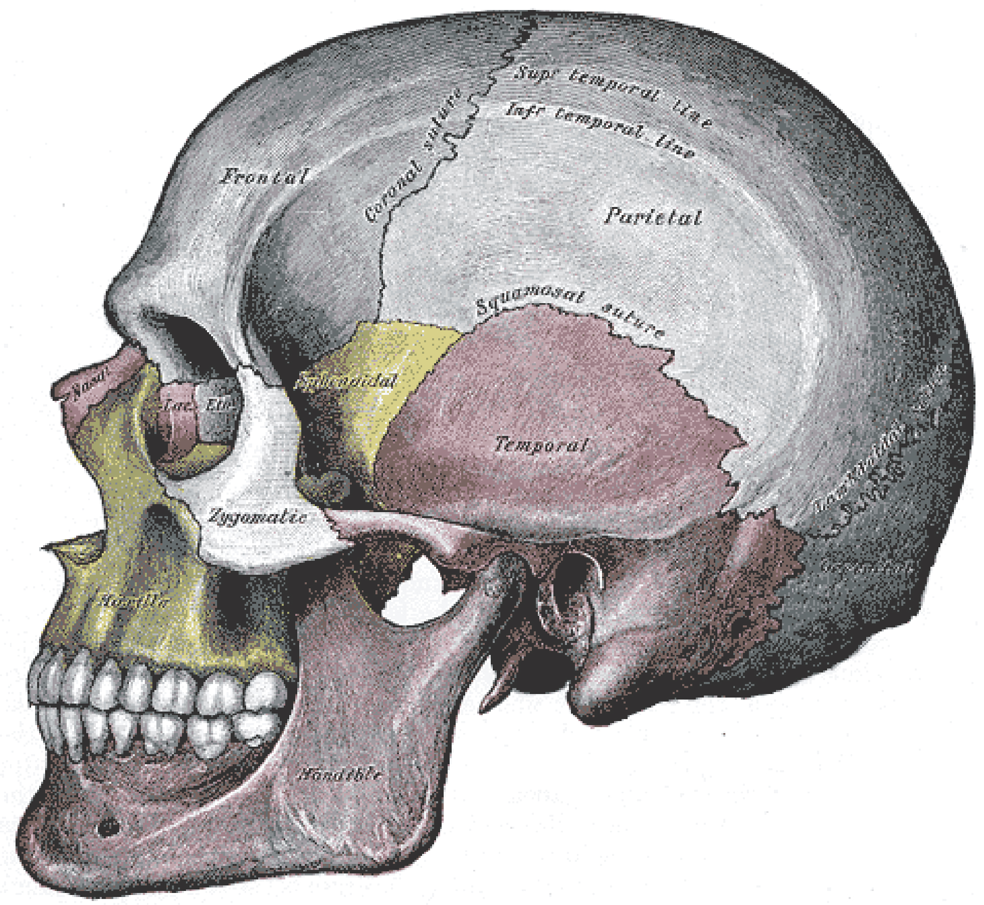
Parietal bone: the main side of the skull. Sphenoid bone: the bone located under the frontal bone, behind the nose and eye cavities. Temporal bone:.

The Bones of the Skull Human Anatomy and Physiology Lab (BSB 141)
Giant cell arteritis (GCA) is a type of vasculitis (blood vessel inflammation) in branches of a large neck artery. A GCA headache is severe and can occur anywhere but is often localized to one side of the head near the temple. Other symptoms include scalp tenderness, vision changes, jaw pain when chewing, and unintended weight loss.; Cervicogenic headache manifests as one-sided pain that.